European Journal of Medical Case Reports
Volume 10(7):236–241
Pulmonary cavitations in poorly controlled diabetic patients: is it tuberculosis? – case series of two patients
Marco Antonio Rodriguez Sanchez1* , Exequiel Aispuro Valenzuela1, Luis David Beltran Ontiveros1, Jose Manuel Aguilar Rubio1, Mariana Denisse Heras Cruz2, Cynthia Nahomi Solis Angulo1, Sofia Myraki Flores Gutierrez1
Correspondence to: Marco Antonio Rodriguez Sanchez
*Department of Internal Medicine, Center for Research and Teaching in Health Sciences, Civil Hospital of Culiacán, Autonomous University of Sinaloa, Culiacán, Mexico.
Email: marcorguez16@gmail.com
Full list of author information is available at the end of the article.
Received: 12 January 2026 | Revised (1): 13 March 2026 | Accepted: 12 April 2026
ABSTRACT
Background:
Poorly controlled type 2 diabetes mellitus (T2DM) increases the risk of severe respiratory infections and pulmonary complications, including lung abscesses and cavitary lesions. In tuberculosis-endemic regions, these findings are often initially attributed to tuberculosis, which may delay the diagnosis and management of other relevant infectious etiologies.
Case Presentation:
We report a case series of two patients with poorly controlled T2DM who developed pulmonary cavitations in the right upper lung lobe following non-tuberculous infectious processes. The first case involved a 22-year-old woman with a recent dengue infection who developed a cavitary pneumonia without signs of sepsis and showed a favorable clinical course with conservative management using intravenous antibiotics. The second case was a 68-year-old man with T2DM and chronic obstructive pulmonary disease, initially treated for community-acquired pneumonia, who subsequently developed severe diabetic ketoacidosis, septic shock, and extensive pulmonary cavitation, requiring intensive care unit admission, broad-spectrum intravenous antibiotics, and surgical lobectomy.
Conclusion:
Pulmonary cavitations in patients with poorly controlled T2DM represent a significant diagnostic and therapeutic challenge, particularly in tuberculosis-endemic settings. This case series underscores the importance of maintaining a high index of suspicion for non-tuberculous etiologies, achieving adequate metabolic control, performing timely imaging studies, and implementing a multidisciplinary approach to prevent adverse outcomes and improve prognosis.
Keywords:
Type 2 diabetes mellitus, tuberculosis, pneumonia, pulmonary cavitation, septic shock.
Background
Poorly controlled type 2 diabetes mellitus (T2DM) is associated with an increased susceptibility to severe respiratory infections, which may manifest as lung abscesses or cavitary lesions [1]. In regions where tuberculosis is endemic, pulmonary cavitations are frequently presumed to be tuberculous in origin, leading to the under recognition of alternative etiologies, partly due to the limited specificity of conventional imaging techniques in differentiating cavitary lung lesions. Although poorly controlled T2DM has been shown to increase both the risk and extent of cavitations secondary to tuberculosis when compared with non-diabetic individuals and those with adequate glycemic control [2], evidence addressing non-tuberculous causes of pulmonary cavitations in this population remains scarce.
In this case series, we describe two patients with poorly controlled T2DM who developed right upper lobe pulmonary cavitations of non-tuberculous origin. These cases highlight the importance of maintaining adequate metabolic control, performing timely and appropriate imaging studies, and implementing a multidisciplinary approach to ensure early diagnosis and optimal management of pulmonary complications in diabetic patients.
Why this case matters
“This case series is clinically relevant because it highlights a common diagnostic error in regions endemic for tuberculosis. Through two contrasting clinical scenarios, this report demonstrates that non-tuberculous infectious etiologies can produce significant pulmonary cavitations in diabetic patients, with markedly different clinical courses and outcomes. The cases underscore the critical role of metabolic control as a determinant of disease severity, progression, and response to treatment. Furthermore, they emphasize the importance of early computed tomography (CT), microbiological confirmation, and individualized management strategies to prevent delays in diagnosis, inappropriate therapies, and potentially preventable complications, such as septic shock, diabetic ketoacidosis (DKA), and the need for surgical intervention. By contributing to the limited literature on non-tuberculous pulmonary cavitations in patients with diabetes, this case series reinforces the need for a high index of suspicion and a multidisciplinary approach to their evaluation and management.“
Case Description
Case 1
A 22-year-old woman with a history of T2DM treated with metformin/liraglutide with suboptimal adherence, morbid obesity (BMI 42 kg/m²), and polycystic ovary syndrome presented with general malaise, retro-orbital headache, and persistent fever lasting 10 days. Dengue infection was confirmed by a positive NS1 antigen test. After an initial clinical improvement, she developed right-sided chest pain, progressive exertional dyspnea, and high-grade fever (39.8°C), prompting her to seek emergency care.
On admission, vital signs were as follows: heart rate 110 bpm, blood pressure 127/63 mmHg, temperature 38.2°C, oxygen saturation 91% on room air, and blood glucose 250 mg/dl. Physical examination revealed generalized petechiae and crackles over the right hemithorax, associated with increased tactile vocal fremitus. Laboratory evaluation showed grade II hypochromic microcytic anemia (Hb 8.9 g/dl), marked leukocytosis (20,640/μl; 85% neutrophils, 20% bands), hyperglycemia, hyponatremia (133 mEq/l), significantly elevated C-reactive protein (150 mg/l), and respiratory alkalosis; additionally, glycated hemoglobin (HbA1c) was requested as part of the diagnostic workup and was reported at 6.8%.
Chest CT revealed a cavitary lesion in the right upper lobe with an air–fluid level, associated with parenchymal consolidation and ipsilateral pleural effusion (Figure 1). Given the patient’s clinical presentation, sputum culture and Gram staining were performed, which demonstrated the presence of yeasts, with isolation of Candida albicans. Additionally, three consecutive sputum samples for acid-fast bacilli smear were reported as negative.
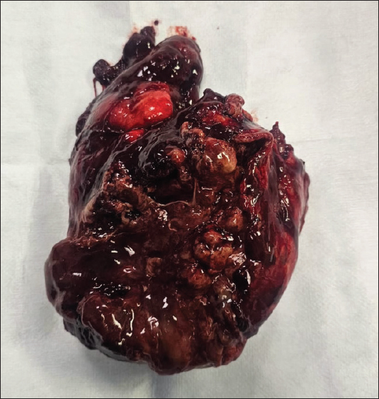
Figure 1. CT of the chest. It shows a cavitary lesion located in the posterior segment of the right upper lobe, with an internal air-fluid level (red arrow), associated with peripheral parenchymal consolidation and inflammatory bronchial changes. Right pleural effusion and mediastinal lymphadenopathy are also observed.
In the absence of sepsis or organ dysfunction (SOFA score 0), conservative management was initiated in a general ward with intravenous ceftriaxone 1 g every 12 hours and metronidazole 500 mg every 6 hours. Furthermore, due to the mycological isolation, antifungal therapy with fluconazole was initiated at 400 mg on the first day, followed by 200 mg every 24 hours thereafter, along with optimization of metabolic control using rapid-acting insulin and close monitoring, resulting in a favorable clinical outcome.
Case 2
A 68-year-old man with a history of T2DM treated with metformin and untreated chronic obstructive pulmonary disease presented with general malaise, productive cough, exertional dyspnea, and fever of 5 days’ duration. Initial chest CT demonstrated consolidation in the right upper lung lobe, leading to a diagnosis of community-acquired pneumonia. Empirical antibiotic therapy was initiated, with initial clinical improvement allowing hospital discharge.
Seventy-two hours after discharge, the patient developed rapidly progressive dyspnea and altered mental status and was readmitted to the emergency department. Severe (DKA; pH 6.80, glucose 397 mg/dl, HCO3− 1.2 mmol/l) and septic shock were diagnosed. On evaluation, oxygen saturation was 74%, with partial improvement after supplemental oxygen at 4 l/min; capillary glucose was 342 mg/dl. Physical examination revealed dry oral mucosa, cold and clammy skin, right-sided thoracic crackles, and delayed capillary refill.
Laboratory tests showed mild normocytic normochromic anemia (Hb 10.1 g/dl), leukocytosis (16,870/μl; 86% neutrophils, 20% bands), acute kidney injury (creatinine 2.0 mg/dl), hypernatremia (155 mEq/l), severe hypokalemia (2.5 mEq/l), and high anion gap metabolic acidosis. Chest CT revealed an extensive cavitary lesion in the right upper lobe (Figure 2). Sputum culture grew Serratia marcescens (40,000 CFU), allowing targeted antimicrobial therapy with piperacillin–tazobactam at a dose of 4.5 g every 8 hours for 8 days. Due to clinical deterioration and lesion extent, the patient required intensive care unit management, broad-spectrum intravenous antibiotics, and surgical intervention with lobectomy; following the lobectomy, the patient was admitted to the intensive care unit and subsequently demonstrated a favorable clinical course (Figure 3).
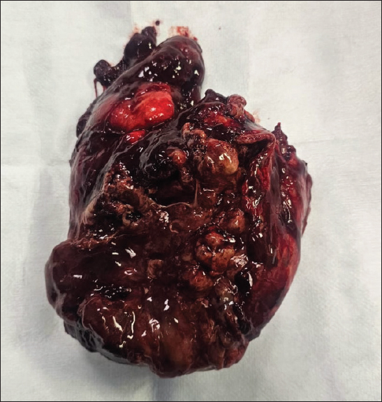
Figure 2. CT of the chest. It reveals an extensive cavitary lesion located in the right upper lobe, containing an air-fluid level, approximately 12 × 9 × 6 cm in diameter (red arrow).
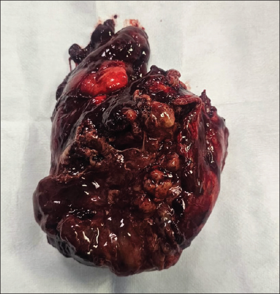
Figure 3. Right pulmonary lobectomy specimen (15 × 13 × 8 cm) showing extensive abscess formation with loss of normal architecture and hemorrhagic areas, consistent with acute necrotizing abscessed pneumonia; no malignancy identified.
Case comparison
Both patients shared poorly controlled T2DM, pulmonary cavitations localized to the right upper lobe, anemia, neutrophilic leukocytosis, and electrolyte and acid–base disturbances, with negative sputum smear microscopy for Mycobacterium tuberculosis. However, marked differences were observed in age, comorbidities, initial clinical presentation, and disease severity. Case 1 involved a young patient without sepsis or organ dysfunction, in whom conservative management was successful. In contrast, Case 2 involved an older patient with severe complications, including DKA and septic shock (SOFA score 7), requiring intensive care management and surgical intervention. These cases highlight the clinical heterogeneity of pulmonary cavitations in patients with poorly controlled T2DM and underscore the importance of individualized evaluation and management strategies.
Discussion
A lung abscess is a serious respiratory infection characterized by a cavitated pulmonary lesion containing purulent material and necrotic tissue [3]. It most commonly develops as a complication of aspiration pneumonia; therefore, aspiration of large volumes of oropharyngeal secretions in patients with impaired consciousness represents a major risk factor. Additional predisposing conditions include structural lung diseases – such as lung malignancies and chronic obstructive pulmonary disease – poor oral hygiene, and immunosuppressive states [4]. Uncontrolled diabetes mellitus causes the patient to be immunocompromised and can be a predisposing factor for other diseases, especially infectious diseases, researchers reported a 50% increase in the risk of Community-acquired pneumonia among patients with Diabetes mellitus (DM) [5].
Pneumonia is common in patients with uncontrolled diabetes mellitus. Based on the literature, hyperglycemia causes an increase in oxidative stress and disturbances in the immune system, like delaying hypersensitivity reactions, lymphocyte transformation, and granuloma formation, which results in changes in immune mechanisms so that DM patients are more susceptible to pneumonia [6].
Lung abscesses are traditionally classified as primary when they result from aspiration of oropharyngeal secretions, and as secondary when they occur in the context of underlying pulmonary disease, bronchial obstruction, or hematogenous dissemination from extrapulmonary infections. Based on their clinical course, abscesses may also be categorized as acute, resolving within 6 weeks, or chronic, persisting beyond this period [7]. In adults, lung abscesses most frequently present between 55 and 75 years of age, with a mean age of approximately 65 years, and show a male predominance, accounting for nearly 65% of cases. The most common symptoms at presentation include cough, malaise, and fever. Lesions typically measure around 5 cm in diameter and are most often located in the right upper or lower lobes [8].
From a diagnostic perspective, the presence of an underlying pulmonary malignancy should always be considered in patients with lung abscesses, as reported prevalence ranges from approximately 7% to higher values depending on the studied population and diagnostic approach [9]. CT remains the gold standard for diagnosis; however, clinical correlation is essential, as differentiating a lung abscess from infectious cysts or other cavitary lung lesions can be challenging. Thoracic ultrasound has emerged as a valuable, accessible imaging modality that provides real-time assessment and may be particularly useful during the initial evaluation [10].
Identification of the causative microorganism is crucial to optimize antimicrobial therapy and limit unnecessary use of broad-spectrum antibiotics; nonetheless, sputum cultures yield positive results in only approximately 50% of cases [11]. In patients who fail to respond to systemic antibiotic therapy, escalation to invasive management should be considered. Factors associated with failure of conservative treatment include endobronchial obstruction due to tumors or foreign bodies, cavitary lesions secondary to malignancy or vasculitis, inappropriate antibiotic selection or insufficient treatment duration, and abscesses larger than 6 cm. In such scenarios, percutaneous transthoracic drainage and endoscopic catheter drainage represent effective alternatives to conventional surgical resection [12].
In the present case series, poorly controlled T2DM played a central role in the development and progression of lung abscesses. Differences in age and comorbidity burden likely explain the variation in disease severity observed between the two patients. Early use of CT for timely diagnosis, prompt initiation of insulin therapy to achieve glycemic control, and appropriate intravenous antibiotic therapy were key factors associated with favorable clinical outcomes. This series contributes to the limited existing literature on lung abscesses in diabetic patients and underscores the importance of a multidisciplinary approach to improve outcomes in this high-risk population.
Conclusion
Pulmonary cavitations in patients with poorly controlled T2DM represent a complex diagnostic and therapeutic challenge, particularly in regions where tuberculosis is endemic and cavitary pulmonary lesions are frequently presumed to be of tuberculous origin. The presentation of these two cases illustrates the clinical spectrum of non-tuberculous pulmonary cavitations in diabetic patients, ranging from favorable outcomes with conservative treatment to severe cases requiring intensive care and surgical intervention. Poor metabolic control, advanced age, and the presence of comorbidities appear to play a fundamental role in disease severity and clinical progression. Early diagnosis, timely imaging acquisition (particularly CT), adequate glycemic control, and targeted antimicrobial therapy are essential to optimize outcomes. A multidisciplinary approach is crucial to prevent diagnostic delays, guide appropriate treatment strategies, and reduce morbidity and mortality in this high-risk population.
What is new?
- This case series highlights that pulmonary cavitations in patients with poorly controlled T2DM may occur following non-tuberculous infectious processes, even in tuberculosis-endemic regions where cavitary lesions are commonly presumed to be tuberculous in origin.
- It illustrates the heterogeneous clinical spectrum of non-tuberculous pulmonary cavitations in diabetic patients, ranging from favorable outcomes with conservative medical management to severe disease complicated by DKA, septic shock, and the need for surgical lobectomy.
- The report emphasizes the central role of poor metabolic control as a key modifier of disease severity, progression, and outcome in pulmonary cavitary infections, independent of patient age or initial clinical presentation.
List of Abbreviations
| CAP | Community-acquired pneumonia |
| COPD | Chronic obstructive pulmonary disease |
| CT | Computed tomography |
| DKA | Diabetic ketoacidosis |
| ICU | Intensive care unit |
| HbA1c | Glycated haemoglobin |
| CRP | C-reactive protein |
| SOFA | Sequential Organ Failure Assessment |
| T2DM | Type 2 diabetes mellitus |
Conflict of interest
The authors declare that they have no conflict of interest regarding the publication of this manuscript.
Funding
This study received no financial support.
Consent for publication
Written informed consent was obtained from the patient for the publication of this case, including clinical information and accompanying images.
Ethical approval
Ethical approval was not required for this case report in accordance with institutional policies; however, all applicable ethical standards and guidelines for research involving human subjects were fully adhered to.
Author details
Marco Antonio Rodriguez Sanchez1, Exequiel Aispuro Valenzuela1, Luis David Beltran Ontiveros1, Jose Manuel Aguilar Rubio1, Mariana Denisse Heras Cruz2, Cynthia Nahomi Solis Angulo1, Sofia Myraki Flores Gutierrez1
- Department of Internal Medicine, Center for Research and Teaching in Health Sciences, Civil Hospital of Culiacán, Autonomous University of Sinaloa, Culiacán, Mexico
- Department of Internal Medicine, Hospital General de Zona No. 2, Mexican Institute of Social Security (IMSS), Autonomous University of Aguascalientes, Aguascalientes, Mexico
References
- Cilloniz C, Torres A. Diabetes mellitus and pneumococcal pneumonia. Diag (Basel). 2024;14(8):859. https://doi.org/10.3390/diagnostics14080859
- Zafar MI, Chen LL, Xiaofeng Y, Gao F. Impact of diabetes mellitus on radiological presentation of pulmonary tuberculosis in otherwise non-immunocompromised patients: a systematic review. Curr Med Imag. 2019;15(6):543–54. https://doi.org/10.2174/1573405614666180806124416
- Sperling S, Dahl VN, Fløe A. Lung abscess: an update on the current knowledge and call for future investigations. Curr Opin Pulm Med. 2024;30(3):229–34. https://doi.org/10.1097/MCP.0000000000001058
- Kuhajda I, Zarogoulidis K, Tsirgogianni K, Tsavlis D, Kioumis I, Kosmidis C, et al. Lung abscess-etiology, diagnostic and treatment options. Ann Transl Med. 2015;3(13):183.
- Kornum JB, Thomsen RW, Riis A, Lervang HH, Schønheyder HC, Sørensen HT. Type 2 diabetes and pneumonia outcomes: a population-based cohort study. Diabetes Care. 2007;30(9):2251–7. doi:10.2337/dc06-2417
- Fahreza F, Novita D, Meutia S. A 56 years old female with uncontrolled diabetes mellitus type II, pneumonia, and suspect with lung abscess: a case report. Jurnal Kedokteran Diponegoro. 2023;12(5):322–5. https://doi.org/10.14710/dmj.v12i5.38079
- Sabbula BR, Rammohan G, Sharma S, Akella J. Lung abscess. Treasure Island, FL: StatPearls Publishing; 2025.
- Vaarst JK, Sperling S, Dahl VN, Fløe A, Laursen CB, Gissel TN, et al. Lung abscess: clinical characteristics of 222 Danish patients diagnosed from 2016 to 2021. Respir Med. 2023;216(107305):107305. https://doi.org/10.1016/j.rmed.2023.107305
- Christie B, A. Umeh C. A lung abscess on CT scan: think of lung cancer. Am J Med Case Rep. 2020;9(1):1–3. https://doi.org/10.12691/ajmcr-9-1-1
- Feki W, Ketata W, Bahloul N, Ayadi H, Yangui I, Kammoun S. Abcès du poumon : diagnostic et prise en charge. Rev Mal Respir. 2019;36(6):707–19. https://doi.org/10.1016/j.rmr.2018.07.010
- Zhang R, Yu J, Shang X, Wang Z, Li H, Cao B. Heterogeneity in clinical patterns of adult lung abscess patients: an 8-year retrospective study in a tertiary hospital. BMC Pulm Med. 2025;25(1):101. https://doi.org/10.1186/s12890-025-03487-2
- Hadid W, Stella GM, Maskey AP, Bechara RI, Islam S. Lung abscess: the non-conservative management: a narrative review. J Thorac Dis. 2024;16(5):3431–40. https://doi.org/10.21037/jtd-23-1561
Summary of the case
| ITEM | CASE 1 | CASE 2 |
|---|---|---|
| Age/sex | 22-year-old female | 68-year-old male |
| Medical history | Poorly controlled T2DM, morbid obesity (BMI 42 kg/m²), polycystic ovary syndrome | Poorly controlled T2DM, chronic obstructive pulmonary disease |
| Presentation | Fever following recent dengue infection, right-sided chest pain, exertional dyspnea | Productive cough, dyspnea, fever; rapid deterioration after initial discharge |
| Key findings | Leukocytosis with neutrophilia and bandemia; anemia; elevated CRP; hyperglycemia; chest CT showing right upper lobe cavitary lesion with air–fluid level and pleural effusion | Severe DKA, septic shock, acute kidney injury; leukocytosis; electrolyte disturbances; chest CT showing extensive right upper lobe pulmonary cavitation |
| Diagnostic workup | Negative sputum smear microscopy for Mycobacterium tuberculosis; CT findings consistent with cavitary pneumonia | Negative sputum smear microscopy for Mycobacterium tuberculosis; sputum culture positive for Serratia marcescens |
| Diagnosis | Non-tuberculous cavitary pneumonia | Lung abscess secondary to Serratia marcescens pneumonia complicated by DKA and septic shock |
| Management | Intravenous antibiotics, optimization of glycemic control, close monitoring in general ward | Intensive care unit admission, insulin infusion, broad-spectrum intravenous antibiotics, electrolyte correction, surgical right upper lobectomy |
| Outcome | Favorable clinical evolution with complete resolution | Clinical improvement after surgical and medical management |
Keywords: Type 2 diabetes mellitus, tuberculosis, pneumonia, pulmonary cavitation, septic shock.


Publication History
Received: January 12, 2026
Revised: March 13, 2026
Accepted: April 12, 2026
Published: June 19, 2026
Authors
Marco Antonio Rodríguez Sánchez
Department of Internal Medicine, Center for Research and Teaching in Health Sciences, Civil Hospital of Culiacán, Autonomous University of Sinaloa, Culiacán, Mexico.
 ORCID
Exequiel Aispuro Valenzuela
Department of Internal Medicine, Center for Research and Teaching in Health Sciences, Civil Hospital of Culiacán, Autonomous University of Sinaloa, Culiacán, Mexico.
Luis David Beltrán Ontiveros
Department of Internal Medicine, Center for Research and Teaching in Health Sciences, Civil Hospital of Culiacán, Autonomous University of Sinaloa, Culiacán, Mexico.
José Manuel Aguilar Rubio
Department of Internal Medicine, Center for Research and Teaching in Health Sciences, Civil Hospital of Culiacán, Autonomous University of Sinaloa, Culiacán, Mexico.
Mariana Denisse Heras Cruz
Department of Internal Medicine, Hospital General de Zona No. 2, Mexican Institute of Social Security (IMSS), Autonomous University of Aguascalientes, Aguascalientes, Mexico.
Cynthia Nahomi Solís Angulo
Department of Internal Medicine, Center for Research and Teaching in Health Sciences, Civil Hospital of Culiacán, Autonomous University of Sinaloa, Culiacán, Mexico.
Sofia Myraki Flores Gutiérrez
Department of Internal Medicine, Center for Research and Teaching in Health Sciences, Civil Hospital of Culiacán, Autonomous University of Sinaloa, Culiacán, Mexico.