European Journal of Medical Case Reports
Volume 10(7):223–228
Double-discoid excision for deep bowel endometriosis – a case report
Yamal Patel1 , Kennedy Malele2*, Charles Muriuki3, Alin Constantin4, Joseph Njagi3
Correspondence to: Kennedy Malele
*Research Department, 3rd Park Hospital, Nairobi, Kenya.
Email: kennedy.malele@gmail.com
Full list of author information is available at the end of the article.
Received: 19 December 2025 | Revised (1): 22 March 2026 | Accepted: 10 April 2026
ABSTRACT
Background:
Deep infiltrating bowel endometriosis (DIE) can result in debilitating pain, dyspareunia, and infertility. Double-discoid full-thickness resection is a fertility-sparing option for selected rectal lesions.
Case Presentation:
A 35-year-old African woman presented with severe dysmenorrhea (VAS 9/10), menorrhagia, dyschezia with alternating constipation and bloating, deep dyspareunia, and infertility. Transvaginal ultrasound demonstrated adenomyosis, myoma, and a 2.72 × 0.78 × 1.75 cm rectal nodule; Enzian classification: A3, B2/2, C2, FA. She underwent diagnostic hysteroscopy with hysteroscopic myomectomy, laparoscopic resection of pelvic endometriosis nodules, adhesiolysis, and staged transanal double-discoid full-thickness excision of the rectal (lesion with a transanal circular stapler. Histopathology of the specimens confirmed endometriosis of the uterosacral and rectal lesions.
Outcome:
The patient was discharged on postoperative day 2, described marked symptomatic improvement at a 10-day follow-up visit, had resumed a normal diet and bowel function, and was started on suppressive hormonal therapy (dienogest). No complications were encountered.
Conclusion:
Double-discoid full-thickness excision of rectal endometriosis is a feasible surgical option for selected rectal DIE nodules <30 mm depth-invasive or when lesion geometry allows; careful patient selection and operative expertise are crucial aspects.
Keywords:
Bowel endometriosis, full-thickness, resection, discoid excision, case report.
Background
Deep infiltrating endometriosis (DIE) of the bowel is a complex clinical diagnosis affecting up to 12% of women with endometriosis [1,2]. Patients often present with severe dysmenorrhea, dyspareunia, dyschezia, bloating, and infertility, symptoms which lead to significant impairment of quality of life [3-5]. Diagnostic delays can be problematic due to overlapping symptoms with other gastrointestinal diseases. High-resolution transvaginal ultrasound and magnetic resonance imaging can provide better preoperative mapping and classification [6-8]. Surgical management for treatment in symptomatic cases is necessary when medical therapy is ineffective at rates [9]. Three main surgical techniques include shaving of seromuscular disease, discoid full-thickness resection, and segmental bowel resection. The selected technique depends on the size, depth, and circumference of the lesion [8-12]. Ultimately, individualized treatment decisions should determine the radicality of excision weighed against the risk of complications such as bowel dysfunction, fistula formation, or anastomotic leakage [13-15]. The discoid excision approach has been widely used to treat rectal DIE in women with fertility-preserving desire. However, what is not as widely applied is the double-discoid excision approach. This report describes the use of a double-discoid full-thickness excision performed transanally in a patient with a longitudinally extensive anterior rectal deep endometriosis lesion that could not be optimally excised with a single stapler, yet had limited circumferential involvement. The decision to complete a full-thickness excision was based on the geometry of the lesion, avoidance of segmental resection, and fertility-preserving desire.
Case Presentation
A 35-year-old African female diagnosed with longstanding severe dysmenorrhea, menorrhagia, dyschezia, alternating constipation and bloating, deep dyspareunia, and infertility. She rated her menstrual pain a 9/10 and also reported intermittent non-cyclic pelvic pain. On pelvic examination, she had reproducible posterior pelvic tenderness; no prior adnexal mass was palpated on bimanual exam. Transvaginal ultrasound for DIE demonstrated adenomyosis, uterine myoma, and a hypoechoic anterior rectal wall nodule measuring 2.72 × 0.78 × 1.75 cm (Figure 1). #Enzian(u): A3, B2/2, C2, FA; intraoperative Enzian(s): T2/2, A2, B2/2, C3, FA. The preoperative and intraoperative evaluation indicated a single anterior rectal DIE nodule with full-thickness muscularis involvement, <50% circumferential involvement, and longitudinal spread greater than the diameter of the staple disc. The depth of the lesion could not allow for complete excision by rectal shaving. Bowel resection was unnecessary, given the absence of disease multifocality, limited circumferential involvement, and rectosigmoid stenosis. Therefore, a staged transanal double-discoid full-thickness excision was considered to achieve complete lesion clearance while providing preservation of fertility and bowel continuity. Differential diagnoses considered included colorectal neoplasm and inflammatory bowel disease, but imaging and operative findings supported DIE. Histopathology confirmed endometriosis in rectal and uterosacral specimens. No history of prior medical therapy for endometriosis was provided.
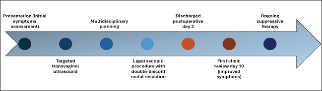
Figure 1. A 2D Sonographic imaging showing the hypoechoic, irregularly contoured rectal nodule measuring 2.72 × 0.8 × 1.75 cm, located in the anterior rectal wall. The lesion appears infiltrative with posterior shadowing, consistent with deep endometriosis.
Surgical approach (step-by-step)
Under laparoscopic guidance, extensive pelvic adhesiolysis, hysteroscopic myomectomy, and laparoscopic shaving of uterosacral nodules were performed. The rectal lesion was mobilized and shaved (Figure 2); angle sutures and two Prolene traction sutures were placed. A transanal circular end-to-end stapler was used for the first full-thickness discoid excision. Due to the longitudinal length of the lesion Figure 4 exceeding the capture capability of a single circular stapler disc, the staged discoid excision provided complete full thickness resection of the DIE lesion while maintaining the integrity of the staple line and bowel continuity (Figure 3). On-table rectoscopy showed the staple line was intact. Two-angle sutures were placed to augment the anastomosis. There were no intraoperative complications. Post-operative analgesics and antibiotics were provided prophylactically as per institutional protocol; no post-operative therapeutic antibiotics were required.
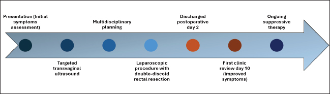
Figure 2. Laparoscopic rectal shaving to excise DIE tissue. The dissection is undertaken carefully down to the anterior rectal wall in the muscularis layer to allow for maximal excision of fibrotic and infiltrative disease while maintaining rectal viability. This is an important step before full thickness rectal disc excision for deeper infiltration.
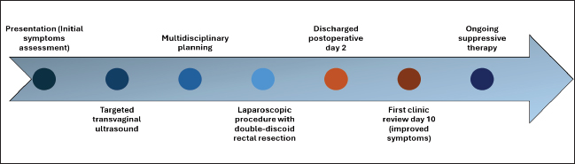
Figure 3. Rectal shaved area caught between the anvil and the shoulder of the transanal circular stapler, which is progressively closed under laparoscopic control.
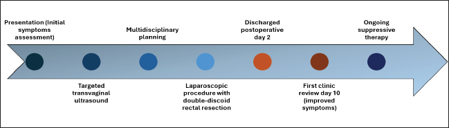
Figure 4. Intraoperative images showing the gross appearance of the resected rectal nodule, with a firm, irregular, fibrotic mass invading the muscularis propria of the anterior rectal wall, consistent with DIE. The surface is nodular and heterogenous and shows signs of fibrosis, and possibly glandular components.
The patient was started on suppressive hormonal therapy with dienogest (2 mg). She was vitally stable, tolerated diet well, and passed stool and urine without complication. At the initial clinic review day 10, the patient stated her symptoms of dyschezia and ≤bloating had almost significantly resolved; she had a significant decrease in her dysmenorrhea (before surgery, her Visual Analogue Scale (VAS) score was 9); and she had normalized bowel function. There were no immediate adverse or unexpected events documented. She was continued on suppressive hormonal therapy with dienogest. Postoperative pain improvement was evaluated based on the patient-reported outcome on a VAS scale. No specific validated quality-of-life assessment instrument was used.
Prior to presentation, the patient described a multiple-year history of progressively worsening cyclical pelvic pain and bowel symptoms. Diagnostic transvaginal ultrasound for DIE mapping was performed, followed by surgical intervention after multidisciplinary discussion. Surgery consisted of a simultaneous laparoscopic and transanal double-discoid excision. The patient was discharged 2 days postoperatively, with a follow up appointment at 10 days, and began suppressive hormonal therapy (Dienogest) after that date Figure 5.
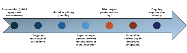
Figure 5. Timeline of the patient’s endometriosis management pathway.
Discussion
This case demonstrates that double-discoid full-thickness excision can be a safe and efficacious fertility-sparing approach to bowel DIE. It highlights the application of a staged double-discoid excision for rectal DIE lesions with unique longitudinal geometry. While discoid excisions have been performed before, this case expands the understanding of the applicability of double-discoid excisions, where a discoid excision or a segmental resection can be undertaken, given the length of the lesion involved. The double-discoid excision in this case preserved continuity of the bowel and the fertility potential of a young woman who was infertile. The patient’s fertility outcomes will continue to be tracked during the patient’s ongoing clinical follow-up; however, there are no pregnancy outcomes available at this time.
The patient had presented with severe cyclical pain and bowel symptoms, which improved significantly after surgery, with bowel function normalizing and no immediate complications. Current literature supports some form of surgical approach for rectal DIE, including shaving, discoid excision, and segmental resections, noting that the type of surgery is dependent on the size of the lesion, depth of lesion, as well as circumferential involvement of the lesion [1-4]. Segmental bowel resection was an option in pre-operative planning, but was unsuitable due to the absence of bowel stenosis, limited circumferential involvement, and the fertility-preserving goal. Wide segmental resection is usually reserved for multiple lesions involving >50% of the circumference or considerable bowel-compromising luminal issues. A double discoid technique offers complete excision of extensive longitudinal lesions with less morbidity associated, in comparison, and is recommended for nodules less than 3 cm and with less than 50% circumferential involvement of the rectum, as in this case [5,6]. Comparatively, general data suggests that long-term pain relief is comparable between discoid and segmental resection; however, discoid approaches offer less morbidity and better functional outcomes than segmental resections [7-9]. Double-discoid approaches provide an option for excising a nodule longer than the reach of a single stapler disc to allow for adequate excision while maintaining some degree of bowel continuity [14,15]. The double-discoid excision technique is generally associated with fewer occurrences of anastomotic leak, post-operative bowel dysfunction, and long-term stenosis when performed in patients selected appropriately, when compared to segmental colorectal resection. Segmental resections may be necessary for extensive or circumferential disease, but the technique has a greater risk of disrupting continuity of the bowel and nerve injury. The double-discoid approach appears to provide a balance between disease excision and a lower incidence of complications associated with the surgery [7-9,14,15].
This case illustrates technical feasibility, multidisciplinary surgical consideration, and documents early symptomatic resolution. The case asserts that double-discoid excision is an acceptable and fertility-sparing approach in selected patients, specifically those with rectal DIE. Good outcomes depend on careful patient selection, advanced laparoscopic skills, and an optimal surgical and medical management approach. The use of dienogest post-operatively is also consistent with the literature that supports medical suppression of disease [12,13]. A significant limitation includes a significantly short duration of follow up; thus, no long-term functional (reproductive) or recurrence outcomes can be assessed at present. The consideration for the preservation of fertility was an influence on the surgical decision-making process; however, pregnancy outcomes were still not available at the time of this report.
Conclusion
The double-discoid excision technique is a feasible operative approach for the treatment of bowel DIE. It allows full excision of endometriotic lesions while preserving bowel continuity in symptomatically troubled patients. This case report provided an example of successful double disc excision in a patient with DIE, with satisfactory post-operative results and improvement in the patient’s symptomatology. The importance of patient selection, surgical experience to perform the surgery, and a multidisciplinary working relationship will ensure the best possible outcomes for the patient.
What is new?
DIE of the bowel is a complicated condition that causes debilitating pelvic pain, bowel symptoms, and can lead to infertility; treatment options include shaving, discoid excision, or segmental resection, depending on the lesion characteristics. Discoid full-thickness excision is a fertility-sparing approach for small rectal nodules. This paper details a new adaptation of a double-discoid (or double-disc) full-thickness excision for rectal DIE. The authors describe the technical feasibility, safety, and good short-term outcomes. Careful patient selection and multidisciplinary surgical planning are crucial aspects.
List of Abbreviations
| DIE | Deep infiltrating endometriosis |
| VAS | Visual Analogue Scale |
Conflicts of interest
The authors declare that they have no conflict of interest regarding the publication of this case report.
Funding
No funding was received from the public, commercial, or not-for-profit sector.
Consent for publication
Written informed consent was obtained from the patient for publication of this case report and any accompanying images. A copy of the written consent is available for review by the Editor-in-Chief of this journal.
Ethical approval
Ethical approval for this case report was obtained from Kenyatta University Ethics Review Committee (KUERC Ref. No.: PKU/3389/14110) dated on:11th November 2025.
Availability of data and material
The data supporting the findings of this case report are not publicly available due to privacy and ethical considerations.
Author contributions
All authors contributed to conception, data collection, manuscript drafting, and final approval of the submitted version.
Author details
Yamal Patel1, Kennedy Malele2, Charles Muriuki3, Alin Constantin4, Joseph Njagi3
- Consultant Obstetrician and Gynaecologist, Hysteroscopic and Laparoscopic Surgeon, 3rd Park Hospital, Nairobi, Kenya
- Research Department, 3rd Park Hospital, Nairobi, Kenya
- Consultant Obstetrician, Gynaecologist and Laparoscopic Surgeon, 3rd Park Hospital, Nairobi, Kenya
- Alin Constantin, Department of Gynecology, Obstetrics and Reproductive Medicine, Saarland University Hospital, Homburg, Germany
References
- Protopapas A, Giannoulis G, Chatzipapas I, Athanasiou S, Grigoriadis T, Haidopoulos D, et al. Posterior deep infiltrating endometriotic nodules: operative considerations according to lesion size, location, and geometry, during One’s Learning Curve. ISRN Obstet Gynecol. 2014;2014:853902. https://doi.org/10.1155/2014/853902
- Donnez O, Roman H. Choosing the right surgical technique for deep endometriosis: shaving, disc excision, or bowel resection?. Fertil Steril. 2017;108(6):931–42. https://doi.org/10.1016/j.fertnstert.2017.09.006
- Ceccaroni M, Ceccarello M, Clarizia R, Fusco E, Roviglione G, Mautone D, et al. Nerve-sparing laparoscopic disc excision of deep endometriosis involving the bowel: a single-center experience on 371 consecutives cases. Surg Endosc. 2021;35(11):5991–6000. https://doi.org/10.1007/s00464-020-08084-4
- Laganà AS, Vitale SG, Trovato MA, Palmara VI, Rapisarda AMC, Granese R, et al. Full-thickness excision versus shaving by laparoscopy for intestinal deep infiltrating endometriosis: rationale and potential treatment options. BioMed Res Int. 2016;2016:3617179. https://doi.org/10.1155/2016/3617179
- Namazov A, Kathurusinghe S, Marabha J, Merlot B, Forestier D, Hennetier C, et al. Double disk excision of large deep endometriosis nodules infiltrating the low and mid rectum: a pilot study of 20 cases. J Minim Invasive Gynecol. 2020;27(7):1482–9. https://doi.org/10.1016/j.jmig.2020.04.019
- Roman H, Dennis T, Forestier D, François MO, Assenat V, Tuech JJ, et al. Disk excision using end-to-end anastomosis circular stapler for deep endometriosis of the rectum: a 492-patient continuous prospective series. J Minim Invasive Gynecol. 2023;30(2):122–30. https://doi.org/10.1016/j.jmig.2022.10.009
- Roman H, Abo C, Huet E, Bridoux V, Auber M, Oden S, et al. Full-thickness disc excision in deep endometriotic nodules of the rectum: a prospective cohort. Dis Colon Rectum. 2015;58(10):957–66. https://doi.org/10.1097/DCR.0000000000000447
- Roman H, Tuech JJ, Huet E, Bridoux V, Khalil H, Hennetier C, et al. Excision versus colorectal resection in deep endometriosis infiltrating the rectum: 5-year follow-up of patients enrolled in a randomized controlled trial. Hum Reprod. 2019;34(12):2362–71. https://doi.org/10.1093/humrep/dez217
- Warring SK, Cope AG, Youssef Y, Vanburen WM, Burnett TL, Langstraat CL, et al. Excision of deep endometriosis of the rectosigmoid: individualizing care to the presenting pathology. J Minim Invasive Gynecol. 2022;29(9):1037. https://doi.org/10.1016/j.jmig.2022.06.017
- Braund S, Hennetier C, Klapczynski C, Scattarelli A, Coget J, Bridoux V, et al. Risk of postoperative stenosis after segmental resection versus disk excision for deep endometriosis infiltrating the rectosigmoid: a retrospective study. J Minim Invasive Gynecol. 2021;28(1):50–6. https://doi.org/10.1016/j.jmig.2020.04.034
- Kathopoulis N, Vlachos DE, Kypriotis K, Diakosavvas M, Chatzipapas I, Protopapas A. Laparoscopic discoid excision of bowel endometriosis using sutures for closure. J Minim Invasive Gynecol. 2023;30(1):11–2. https://doi.org/10.1016/j.jmig.2022.11.007
- Roman H. Disc excision using transanal circular stapler for deep endometriosis of the rectum in 10 steps. J Minim Invasive Gynecol. 2021;28(1):14–5. https://doi.org/10.1016/j.jmig.2020.04.017
- Donnez O. Conservative management of rectovaginal deep endometriosis: shaving should be considered as the primary surgical approach in a high majority of cases. J Clin Med. 2021;10(21):5183. https://doi.org/10.3390/jcm10215183
- Pinho Oliveira MA, Crispi CP, Oliveira FM, Reis PS, Raymundo TS, Pereira TD. Double circular stapler, or laparoscopic double discoid resection with a circular stapler. J Minim Invasive Gynecol. 2016;23(5):844. https://doi.org/10.1016/j.jmig.2016.01.031
- Kondo W, Ribeiro R, Zomer MT, Hayashi R. Laparoscopic double discoid resection with a circular stapler for bowel endometriosis. J Minim Invasive Gynecol. 2015;22(6):929–31. https://doi.org/10.1016/j.jmig.2015.04.021
Summary of case
| 1 | Patient (gender, age) | 35 years, female |
| 2 | Final diagnosis | Deep infiltrating rectal endometriosis |
| 3 | Symptoms | Severe dysmenorrhea (VAS 9/10), menorrhagia, dyschezia, constipation and bloating, deep dyspareunia, and infertility |
| 4 | Medications | Postoperative suppressive hormonal therapy - dienogest (2 mg daily); perioperative analgesics and antibiotics |
| 5 | Clinical procedure | Laparoscopic + transanal double-disc full-thickness excision of endometriosis |
| 6 | Specialty | Obstetrics and gynaecology – minimally invasive / laparoscopic surgery |
Keywords: Bowel endometriosis, full-thickness, resection, discoid excision, case report.


Publication History
Received: December 19, 2025
Revised: March 22, 2026
Accepted: April 10, 2026
Published: June 19, 2026
Authors
Yamal Patel
Consultant Obstetrician and Gynaecologist, Hysteroscopic and Laparoscopic Surgeon, 3rd Park Hospital, Nairobi, Kenya.
 ORCID
Kennedy Malele
Research Department, 3rd Park Hospital, Nairobi, Kenya.
Charles Muriuki
Consultant Obstetrician, Gynaecologist and Laparoscopic Surgeon, 3rd Park Hospital, Nairobi, Kenya.
Alin Constantin
Alin Constantin, Department of Gynecology, Obstetrics and Reproductive Medicine, Saarland University Hospital, Homburg, Germany.
Joseph Njagi
Consultant Obstetrician, Gynaecologist and Laparoscopic Surgeon, 3rd Park Hospital, Nairobi, Kenya.