European Journal of Medical Case Reports
Volume 10(5):130–135
Rheumatoid arthritis diagnostic pitfalls: don’t overlook large joint effusion - a case report
Perdana Aditya Rahman1 , Michelle Anabel2*, Dyto Prasetya Buwana2, Farah Diyah Anjani2
Correspondence to: Michelle Anabel
*Faculty of Medicine, Brawijaya University, Malang, Indonesia.
Email: michelleanabellll@gmail.com
Full list of author information is available at the end of the article.
Received: 09 December 2025 | Revised (1): 17 February 2026 | Accepted: 18 February 2026
ABSTRACT
Background:
Rheumatoid arthritis (RA) is a systemic autoimmune disease that commonly presents as inflammatory symmetric polyarthritis in small joints and may also have extra-articular involvement. Early diagnosis and treatment within the first 3 months are important to reduce progression and deformities. In some cases, large joint involvement may also present or initially present in RA, which may lead to lower suspicion of RA at the initial visit and result in delayed diagnosis.
Case Presentation:
Here, we present a case of female, 55 years old with knee joint pain and effusion, which was initially thought to be osteoarthritis. The patient had inflammatory arthritis features, including morning stiffness, pain worsened at rest, and arthritis also involving hands. Yet, the patient was confirmed as having rheumatoid arthritis based on clinical, serological, and radiological features. In the detailed evaluation of knee radiographic examination, bone erosions were identified at a second look.
Conclusion:
In cases of bilateral large joint involvement, radiological evaluation should be carefully considered, as rheumatoid arthritis may mimic and superimpose degenerative joint disease, considered as atypical degenerative findings. A comprehensive clinical assessment supported by serological and radiological findings is essential to avoid delayed diagnosis and enable timely treatment.
Keywords:
Knee joint, joint effusion, joint disease, rheumatoid arthritis, osteoarthritis, case report.
Introduction
Knee joint involvement might be a diagnostic pitfall in the diagnosis of inflammatory arthritis, facing the trends that RA prevalence increases with age and knee joint involvement is classically considered as osteoarthritis, in which knee Osteoarthritis (OA) is the most prevalent among other sites [1,2]. Typical clinical manifestations of RA are symmetrical inflammatory polyarthritis of small joints, especially interphalangeal proximal joints. But on further progression, there can be large joint involvement, such as wrist, elbow, shoulder, knee, ankle, and hip joint [3]. In a 2004 study in Korea, the prevalence of RA in knee joints was 42%, the third large joint following the wrist and ankle [4].
The most common radiographic feature of RA is joint erosion, which is also found in psoriatic arthritis [5,6]. Recent classification criteria for RA exclude radiographic features since the presence of radiographic erosions is found in the progressive phase, while earlier diagnosis is required to achieve better outcomes. The last criterion, including radiographic features, was American College of Rheumatology (ACR) 1987.
Case Presentation
A 55-year-old female with a body mass index of 31 kg/m2 was admitted to the emergency unit complaining of worsening pain in both knees lasting for the last 2 weeks. Knee pain has been recurrent for the past 6 months, worsening with activity, especially when raised after sitting, lasting for an hour. The pain worsened and was accompanied by stiffness. Further history-taking reveals other joint involvement, including wrists, fingers, and morning stiffness. On physical examination, we found swelling and tenderness accompanied by a limited range of motion of both knees (Figure S2-S4), also tenderness on right PIP II–III, left PIP II– IV, and left MCP II (Figure S1). Knee Radiographic examination was initially assessed as tibiofemoral osteoarthritis K-L grade IV, with soft tissue swelling and suspected joint effusion.
Table 1. Laboratory Findings.
| Complete blood count | ||
| Hemoglobin | 9,90 g/dl | 13,2-17,3 |
| Erythrocyte | 4,01 × 10^6 | 4,0-5,5 |
| Leucocyte | 17,73 × 10^3 | 4,3-10,3 |
| Hematocrit | 33,2% | 40,0-47,0 |
| Thrombocyte | 536 × 10^3 | 142-424 |
| Differential count |
0,5/0,4/82,2/ |
0-4/0,0-1,0/51-67/ |
| Synovial fluid analysis result | ||
| Erythrocyte | 4,992,1 /µl | |
| Leucocyte | 2,301,6 /µl | |
| PMN cell | 91,0% | |
| MN cell | 9,0% | |
| Crystal | Negative | |
| Serology and autoimmune analysis result | ||
| Rheumatoid factor total | 150,00 IU/ml | < 14 |
| Anti CCP 3 | 237,1 U/ml | < 20 |
Due to joint effusion findings, arthrocentesis was further performed for both knees. Joint effusion of cloudy yellow joint fluid and clouds were obtained, and the microscopic shows typical inflammatory synovial fluid (Table 1). This findings, alongside with the clinical features and anaemia on laboratory findings rise the suspicion of autoimmune arthritis, therefore the patient was consult to the rheumatology department. Reviewing the radiographic findings, marginal erosions were also identified on the medial aspects of the right tibia and lateral aspects of left femur and tibia. Giving the patient characteristics, female, over 50 years old, obese, and initial radiographic finding, it is common in clinical settings that the patient was considered as osteoarthritic. Yet, small joint involvement (even large joints experienced more prominent inflammation) should always be consideration of inflammatory arthritis including rheumatoid arthritis.
Further serologic testing showing high positive anti-CCP3 and rheumatoid factor. Then, based on ACR European League Against Rheumatism (EULAR) Classification Criteria for Rheumatoid Arthritis score obtained is 10 (including: >10 joints, high positive rheumatoid factor /Anti–Cyclic Citrullinated Peptide Antibody, abnormal C-Reactive Protein / Erythrocyte sedimentation rate, complaint duration ≥6 weeks), the patient was classified as rheumatoid arthritis. Patient then commenced treatment with methotrexate 7.5 mg weekly.
Discussion
Rheumatoid arthritis predominantly involves small joints of the hands and feet because they contain abundant synovial tissue, the primary target of autoimmune inflammation [7]. Their complex structure and rich vascular supply make them especially susceptible to the early, chronic, and symmetric inflammatory process characteristic of RA. In the 2010 ACR/EULAR classification criteria, the number and distribution of involved joints are critical for achieving a score of ≥6, which is required to classify a patient as having definite RA. In particular, involvement of small joints - such as the metacarpophalangeal (MCP), proximal interphalangeal (PIP), and wrist joints - contributes more significantly to the overall score than involvement of large joints alone, reflecting the typical pattern of early disease presentation [8]. As the disease progresses, larger joints may become involved, most commonly the wrist, followed by the ankle and knee [3]. The prevalence of knee involvement in RA patients varies, ranging from 30% to 50% at the initial phase and 70%-80% as the disease progresses, where these findings are more frequent in men [9,10]
A study by Abdel-Tawab, et al. [11] taken place in Egypt show that the knee is the sixth most involved joint after MCP, PIP, wrist, Metatarsophalangeal, and Temporomandibular Joint (TMJ). This shows that aside from the hand and wrist, the distribution of joint involvement between regions may vary. In Indonesia, TMJ is not prevalent, as is the case in Turkey, resulting into lesser clinical suspicion of RA when TMJ is involved in these regions [12].
RA and OA differ fundamentally in their mechanisms of joint damage, and these differences are clearly reflected in radiographic findings. In rheumatoid arthritis, chronic autoimmune activation leads to synovial hyperplasia and pannus formation - an invasive inflammatory tissue that erodes cartilage and penetrates subchondral bone. Pro-inflammatory cytokines such as Tumor Necrosis Factor-alpha, Interleukin 1, and Interleukin 6 promote angiogenesis and upregulate Receptor Activator of Nuclear Factor kappa-B Ligand, stimulating osteoclast-mediated bone resorption. As a result, plain radiographs typically show marginal erosions, particularly at the “bare areas” of the joint, along with periarticular osteopenia due to hyperemia and inflammatory bone loss. Joint space narrowing is usually symmetric, reflecting uniform cartilage destruction from persistent synovitis. In advanced disease, deformities and subluxations may develop due to ligamentous and capsular damage [13].
In contrast, osteoarthritis is driven by mechanical stress (overload) and progressive cartilage degeneration. Cartilage loss exposes subchondral bone, triggering a reparative response mediated by growth factors such as Transforming Growth Factor-beta. This leads to chondrocyte proliferation and endochondral ossification at joint margins [14]. Radiographically, OA is characterized by osteophyte formation, subchondral sclerosis, and sometimes subchondral cysts. Joint space narrowing is typically asymmetric, corresponding to uneven mechanical loading. Unlike RA, erosions are not a dominant feature; instead, imaging reflects bone proliferation and remodeling rather than inflammatory destruction [15].
Presenting effusion could also be evaluated through lateral knee radiography, showing an increase in soft tissue density on the suprapatellar recess. In large effusion volume, anterior displacement from the posterior margin of the quadriceps tendon could be evaluated Llopis et al. [16]. Even so, knee joint involvement in osteoarthritis is a common finding due to its weight-bearing nature. But, RA may also present with knee joint involvement even at the earlier phase.
In our patient, the anteroposterior X-ray of the right knee reveals genu erosion without osteophyte formation. Joint space narrowing is observed in all three compartments, with the most severe narrowing present in the lateral (non-weight-bearing) compartment (Figure 1A). Meanwhile, lateral X-ray imaging of the right knee shows an increasing density of the suprapatellar recess, marking the presence of effusion (Figure 1C). The patient subsequently underwent joint fluid aspiration, with the result of leukocytes of 2.301 cells/ml, with 91% polymorphonuclear cell (PMN), showing an inflammatory effusion.
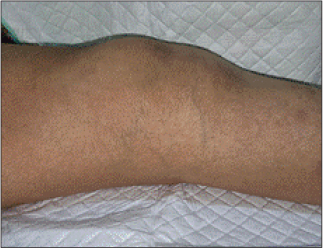
Figure 1. Knee radiographics A. Right knee AP position, B. Left knee AP position, C. Right knee lateral position, D. Left knee lateral position. In Figures A and B, the white arrow shows marginal erosion, blue arrow shows joint space narrowing, and the purple arrow shows minimal osteophyte formation. In figures C and D, the white arrow shows increasing density of soft tissue on the suprapatellar recess.
In the patient’s left knee, joint effusion was observed, along with radiographic findings of erosion and joint space narrowing on the anteroposterior view (Figure 1B and 1D). However, compared to the right knee, the left knee also showed osteophyte formation and predominant joint space narrowing in the medial (weight-bearing) compartment. These findings suggest the possibility of secondary osteoarthritis superimposed on rheumatoid arthritis in the left knee [17].
Conclusion
This case underscores a key diagnostic pitfall in rheumatoid arthritis: delayed recognition due to underappreciation of radiographic erosions in large joints. Although the initial presentation with knee effusions mimicked osteoarthritis, the presence of marginal erosions, minimal osteophyte formation, and symmetric compartmental involvement were atypical for degenerative disease and suggestive of inflammatory arthritis. Failure to recognize these radiographic red flags can postpone appropriate investigation and management. An integrated assessment - combining clinical findings, inflammatory synovial fluid analysis, serologic markers, and characteristic imaging features - is essential to prevent delayed diagnosis, particularly in resource-limited settings.
What is new?
Rheumatoid arthritis is a chronic autoimmune disease that primarily affects small joints, typically presenting with marginal erosions, soft tissue swelling, osteoporosis, and joint space narrowing. It is well known for causing progressive joint destruction, especially in the hands and wrists. What is new in this manuscript is that, although less commonly observed, the radiologic features of rheumatoid arthritis in large joints can overlap with those of osteoarthritis, which may lead to potential misdiagnosis. In cases of knee joint (RA genu) involvement, radiographic findings may include joint effusion, joint space narrowing in the non–weight-bearing area, marginal erosions, and juxta-articular osteopenia.
Acknowledgment
The authors would like to thank Achmad Bayhaqi for his valuable input and expertise in radiology.
List of Abbreviations
| ACR | American College of Rheumatology |
| Anti-CCP | Anti–Cyclic Citrullinated Peptide Antibody |
| AP | Anteroposterior |
| CBC | Complete blood count |
| CRP | C-Reactive Protein |
| Eo | Eosinophil |
| ESR | Erythrocyte Sedimentation Rate |
| EULAR | European League Against Rheumatism |
| IL-1 | Interleukin 1 |
| IL-6 | Interleukin 6 |
| K–L | Kellgren–Lawrence (grading system) |
| Lym | Lymphocyte |
| MCP | Metacarpophalangeal (joint) |
| MN | Mononuclear cell |
| Mon | Monocyte |
| MTP | Metatarsophalangeal (joint) |
| Neu | Neutrophil |
| OA | Osteoarthritis |
| PIP | Proximal Interphalangeal (joint) |
| PMN | Polymorphonuclear Cell |
| RA | Rheumatoid Arthritis |
| RANKL | Receptor Activator of Nuclear Factor kappa-B Ligand |
| RF | Rheumatoid factor |
| ROM | Range of motion |
| TGF-β | Transforming growth factor-beta |
| TMJ | Temporomandibular Joint |
| TNF-α | Tumor necrosis factor-alpha |
Conflict of interest
The authors declare that there is no conflict of interest regarding the publication of this article.
Consent to participate
Written informed consent was obtained from the patient.
Funding
None.
Ethical approval
Ethical approval is not required at our institution for anonymous case reports.
Author details
Perdana Aditya Rahman1, Michelle Anabel2, Dyto Prasetya Buwana2, Farah Diyah Anjani2
- Rheumatology-Immunology Division, Department of Internal Medicine, Faculty of Medicine, Brawijaya University, Dr. Saiful Anwar Hospital, Malang, Indonesia
- Faculty of Medicine, Brawijaya University, Malang, Indonesia
References
- Zhang Z, Gao X, Liu S, Wang Q, Wang Y, Hou S, et al. Global, regional, and national epidemiology of rheumatoid arthritis among people aged 20-54 years from 1990 to 2021. Sci Rep. 2025;15(1):1–12. https://doi.org/10.1038/s41598-025-92150-1
- Steinmetz JD, Culbreth GT, Haile LM, Rafferty Q, Lo J, Fukutaki KG, et al. GBD 2021 osteoarthritis collaborators. Global, regional, and national burden of osteoarthritis, 1990-2020 and projections to 2050: a systematic analysis for the Global Burden of Disease Study 2021. Lancet Rheumatol. 2023;5(9):e508–22.
- Zhao SS, Nikiphorou E, Young A, Kiely PD. Large joints are progressively involved in rheumatoid arthritis irrespective of rheumatoid factor status-results from the early rheumatoid arthritis study. Rheumatol Int. 2022;42(4):621–9. https://doi.org/10.1007/s00296-021-04931-2
- Yoon BY, Lee CH, Han SH, Lee YW. Clinical characteristics and joint involvement of rheumatoid arthritis. J Korean Rheum Assoc. 2004;11(3):227–34.
- Scherer HU, Häupl T, Burmester GR. The etiology of rheumatoid arthritis. J Autoimmun. 2020;110:102400. https://doi.org/10.1016/j.jaut.2019.102400
- Østergaard M, Boesen M. Imaging in rheumatoid arthritis: the role of magnetic resonance imaging and computed tomography. Radiol Med. 2019;124(11):1128–41. https://doi.org/10.1007/s11547-019-01014-y
- Alivernini S, Firestein GS, McInnes IB. The pathogenesis of rheumatoid arthritis [Internet]. Immunity. 2022;55(12):2255–70. Available from: https://www.sciencedirect.com/science/article/pii/S1074761322005994
- Aletaha D, Neogi T, Silman AJ, Funovits J, Felson DT, Bingham CO 3rd, et al. 2010 rheumatoid arthritis classification criteria: an American College of Rheumatology/European League Against Rheumatism collaborative initiative [Internet]. Ann Rheum Dis. 2010;69(9):1580–8. Available from: https://ard.eular.org/article/S0003-4967%25252824%25252919350-8/fulltext
- Schäfer VS, Schmidt WA, Backhaus M, Hartung W. Arthritis of the knee joint in rheumatoid arthritis - evaluation of treatment response by ultrasound in daily clinical practice. Open Rheumatol J. 2016;10(1):81–7. https://doi.org/10.2174/1874312901610010081
- Kevin Pratama M, Atik N, Hamijoyo L. The pattern of joints involvement in patients with rheumatoid arthritis in rheumatology clinic dr. hasan sadikin general hospital bandung. Indonesian J Rheumatol. 2017;9(2):231050. https://doi.org/10.37275/ijr.v9i2.69
- Abdel-Tawab RR, Bani Sayed DS, Ismail AH. Pattern of rheumatoid arthritis in minia governorate. MJMR. 2020;31(3):1–12. https://doi.org/10.21608/mjmr.2020.219850
- Gao Y, Zhang Y, Liu X. Rheumatoid arthritis: pathogenesis and therapeutic advances [Internet]. MedComm. 2024;5(3):e509. https://doi.org/10.1002/mco2.509
- Stanislavsky A, Campos A, Elfeky M. Rheumatoid arthritis (musculoskeletal manifestations). [cited 2026 Mar 01]. https://doi.org/10.53347/rID-12370
- Shen J, Li S, Chen D. TGF-β signaling and the development of osteoarthritis [Internet]. Bone Res. 2014;2(1):14002. Available from: https://pmc.ncbi.nlm.nih.gov/articles/PMC4274935/
- Mous A, Walizai T, Campos A. Osteoarthritis. [cited 2026 Mar 01]. https://doi.org/10.53347/rID-1794
- Llopis E, Kroon HM, Acosta J, Bloem JL. Conventional radiology in rheumatoid arthritis. Radiol Clin North Am. 2017;55(5):917–41. https://doi.org/10.1016/j.rcl.2017.04.002
- Vad VB, Adin DR, Solomon J. Knee osteoarthritis. Crit Rev Phys Rehabil Med. 2023;16(3):211–31. https://doi.org/10.1615/CritRevPhysRehabilMed.v16.i3.30
Summary of the case
| 1 | Patient (gender, age) | Female, 55 |
| 2 | Final diagnosis | Rheumatoid arthritis (with knee joint involvement) |
| 3 | Symptoms | Bilateral knee pain with swelling and morning stiffness |
| 4 | Medications | Methotrexate 7.5 mg weekly |
| 5 | Clinical procedure | Arthrocentesis |
| 6 | Specialty | Rheumatology |
SUPPLEMENTARY MATERIAL
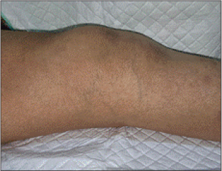
Figure S1. Left Hand. Swelling (+), Boutonniere deformity (+), and ulnar deviation (+).
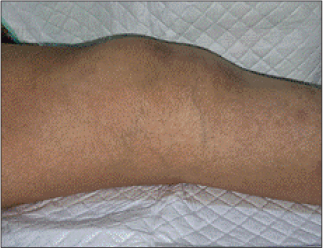
Figure S2. Knee D/S with bilateral swelling.
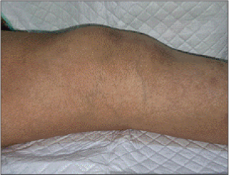
Figure S3. Knee dextra, medial view.
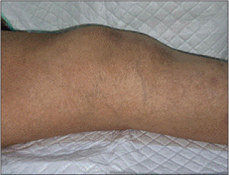
Figure S4. Knee sinistra, medial view.
Keywords: Knee joint, joint effusion, joint disease, rheumatoid arthritis, osteoarthritis, case report.


Publication History
Received: December 09, 2025
Revised: February 17, 2026
Accepted: February 18, 2026
Published: April 13, 2026
Authors
Perdana Aditya Rahman
Rheumatology-Immunology Division, Department of Internal Medicine, Faculty of Medicine, Brawijaya University, Dr. Saiful Anwar Hospital, Malang, Indonesia.
 ORCID
Dyto Prasetya Buwana
Faculty of Medicine, Brawijaya University, Malang, Indonesia.
Farah Diyah Anjani
Faculty of Medicine, Brawijaya University, Malang, Indonesia.