European Journal of Medical Case Reports
Volume 10(8):247–255
Vascular synovial sarcoma masquerading as deep vein thrombosis: a diagnostic
pitfall-a case report
Ritwika Choudhury1 , Bhumi Agarwal2, Ambarish Satwik3, Mansi Sharma2, Abhinav Dewan2, Sabeena K. Choudhary2, Sunil Pasricha4, Himanshu Rohela5, Ullas Batra2*
Correspondence to: Ullas Batra
*Rajiv Gandhi Cancer Institute and Research Centre, Sector 5, Rohini, India.
Email: ullasbatra@gmail.com
Full list of author information is available at the end of the article.
Received: 09 March 2026 | Revised: 25 April 2026 | Accepted: 30 April 2026
ABSTRACT
Background:
Intravascular synovial sarcoma is a rare disease with only 14 cases documented globally to date. To the best of our knowledge, we present the 15th case, highlighting the diagnostic challenge, the rapidly progressive course of this malignancy, and the need for greater clinical awareness.
Case Presentation:
A female patient in her early 20s presented with a painful swelling in the right inguinal region and right lower limb oedema, initially considered to be DVT. Imaging revealed a mass involving the right external iliac and common femoral veins. She underwent surgical excision of the mass, and histopathological evaluation confirmed intravascular biphasic synovial sarcoma, supported by molecular diagnostic testing. The patient received postoperative chemotherapy. However, there was local recurrence of the disease within 5 months of completing chemotherapy and within a year of surgical resection.
Conclusions:
This case emphasizes that not all cases presenting as DVT may be benign, especially in young patients without apparent risk factors. Underlying malignancies, including sarcomas, should be considered in the differential diagnosis. Advanced imaging techniques are critical for early detection and surgical planning. Early multidisciplinary involvement is essential for optimizing outcomes. Given the rarity and aggressive nature of this disease, further research is needed to guide standardized treatment strategies and improve survival.
Keywords:
15th case, intravascular synovial sarcoma, deep vein thrombosis, thromboembolic events, diagnostic challenge
Background
Synovial sarcoma is a rare subtype of soft tissue sarcoma that generally affects young adults. While it has traditionally been associated with the musculoskeletal system, there have been an increasing number of cases with presentation in unusual locations. Amongst these, the rarest is intravascular synovial sarcoma. These tumours often present with non-specific symptoms with features resembling thromboembolic events, making diagnosis challenging. Accurate diagnosis requires a combination of advanced imaging, histopathology, immunohistochemistry, and molecular testing. Due to its rarity, there is limited understanding of the disease’s behaviour, optimal management, and long-term outcomes. We present a case of a young female patient with intravascular synovial sarcoma involving the right external iliac and femoral veins. We aim to add to the limited published data to raise awareness of this unusual presentation and highlight the importance of early suspicion, timely diagnosis, and multidisciplinary care.
Case Presentation
A female of South Asian descent in her early 20s presented with a 2-month history of a swelling in the right inguinal region. It was insidious in onset, gradually increasing in size, associated with progressive worsening of pain and concurrent swelling of the right lower limb. There was no history of trauma to the leg, insect bite, fever, loss of function or sensation of the affected limb, joint swellings, abdominal pain or distension, and any abnormal vaginal discharge or bleeding.
On examination, a large, firm, tender mass measuring approximately 10 × 8 cm was palpable extending from the right iliac fossa toward the umbilicus. The right lower limb showed pitting oedema from the thigh to the ankle, along with warmth, tenderness, and erythema along the deep venous territory. Prominent superficial varicosities were noted over the posterior and lateral aspects of the right thigh, and all distal pulses were palpable. The neurological, systemic, abdominal, and pelvic examinations were unremarkable.
In view of the patient’s presentation, a bilateral lower limb ultrasonography (USG) with venous Doppler was done, which revealed a large, heterogeneously hypoechoic lesion in the right inguinal region ~11.5 × 5.5 cm, containing internal specks of calcification and exerting compressive effects on the right common femoral vein. No definite colour flow or venous spectral waveform was identified, suggesting a deep vein thrombosis (DVT), and features of cellulitis were also observed.
A CT venography of the abdomen and bilateral thighs revealed a soft tissue density mass extending inferiorly into the right thigh and superiorly into the lower pelvis. It exhibited heterogeneous enhancement with internal vascularity and calcific foci, which were seen tracking along the expected anatomical course of the right external iliac and common femoral veins. Multiple right inguinal lymph nodes were seen. These radiological features raised a strong suspicion of an intravascular tumour mimicking the clinical and sonographic appearance of a DVT. A metastatic workup was initiated, and a whole-body PET-CT scan revealed that the disease was localized (Figure 3).
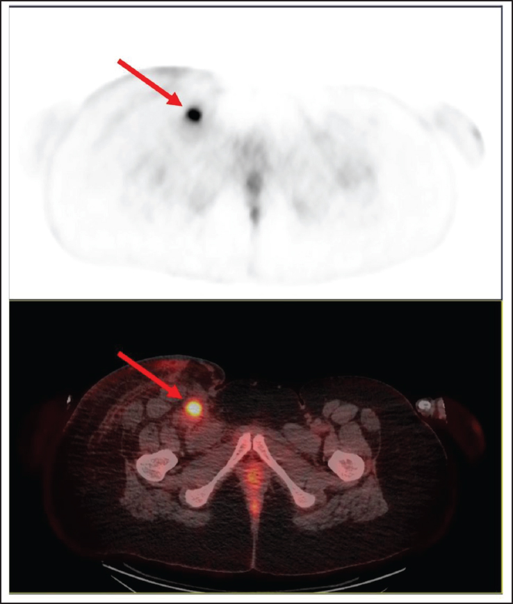
Figure 3. (A–C) No metabolically active lesion seen in right external iliac and inguinal region (post treatment). (D–F) Metabolically active defined soft tissue lesion (yellow arrow) in right inguinal and external iliac region (before treatment).
To establish a definitive diagnosis through histopathological evaluation of the mass, the patient underwent En bloc (surgical removal of the tumour along with surrounding healthy tissue in one single piece) surgical excision of the right iliac mass with excision of the right external iliac vein, right common femoral vein, along with para-aortic, retroperitoneal, and right inguinal lymph nodes dissection. This was followed by vascular reconstruction of both veins using the saphenous vein autograft.
Intraoperatively, a large mass approximately 15 × 10 cm in size was identified arising from the right external iliac vein and common femoral vein. There was significant compression of the right external iliac and common femoral veins, contributing to chronic venous insufficiency. Additionally, there was enlarged right inguinal lymphadenopathy, raising concern for locoregional spread. There was no evidence of DVT in the bilateral lower limb venous systems. This confirmed that the initial diagnosis of DVT was inaccurate and caused by the tumour compression and luminal obstruction.
Grossly, the specimen was a segment of the right iliac vein with attached adipose tissue, measuring 13 × 5 × 4 cm. The external surface appeared congested. On the cut section, a well-circumscribed, encapsulated, grey-white soft to firm nodule measuring 6 × 3.2 × 3 cm was identified. Microscopic evaluation demonstrated a highly cellular, biphasic tumour composed of gland-like epithelial structures interspersed with spindle cell components. Numerous vascular channels were noted, and the tumour was seen expanding within a large vein. Tumour deposits were also present in the surrounding soft tissue. All 19 dissected lymph nodes were free of tumour involvement, and all resected surgical margins were negative for tumour (R0 resection).
Immunohistochemistry (IHC) revealed strong and diffuse SS18 expression in both epithelial and spindle cell components, with CK7 positivity in the epithelial areas. The tumour was negative for SMA, TTF-1, GATA3, PAX8, MYOD1, S100, and CK20. There was significant downregulation of INI1 expression. These findings collectively made the diagnosis of intravascular synovial sarcoma (IVSS), and it was confirmed by the detection of the SYT-SSX1 gene fusion via FISH (Fluorescence in-situ hybridization) analysis on the specimen. Postoperative imaging was unremarkable. There were no postoperative complications.
Following surgical management, the patient received five cycles of chemotherapy (CT) with an ifosfamide and adriamycin regimen. A multidisciplinary discussion with the radiation oncology team was undertaken to discuss the role of local radiotherapy (RT), which was subsequently deferred due to the high risk of chronic right lower limb lymphedema associated with the anatomical site of disease. At 1 and 3 months following the completion of treatment, imaging revealed no evidence of residual or recurrent disease. The patient was kept on regular follow-up.
However, 5 months after completing CT and within 1 year after surgical resection, the patient presented with recurrent swelling of the right lower limb and pain at the postoperative site. MRI and Doppler imaging revealed enhancing multifocal lesions of varying sizes in the subcutaneous and intermuscular planes of the upper right thigh, along with multiple iliac and inguinal region lymph nodes (Figures 1,2). These findings were suggestive of locoregional recurrence. To confirm the diagnosis, a biopsy was performed from the right inguinal subcutaneous lesion, which came back positive for recurrence (Figure 4). A follow-up whole-body PET-CT scan further confirmed the findings without evidence of distant metastasis (Figure 4). The patient was advised to initiate second-line CT with gemcitabine and docetaxel, and Next-Generation Sequencing (NGS) testing of the new lesions to guide further management. Unfortunately, the patient was lost to follow-up thereafter, and no further clinical information is available regarding subsequent management or disease progression.
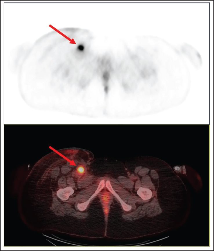
Figure 1. Sagittal STIR image of right upper thigh showing multiple variable sized T2W hyperintense enhancing lesions ssen in subcutaneous and intramuscular planes (yellow arrow) encasing the vessel (red arrow).
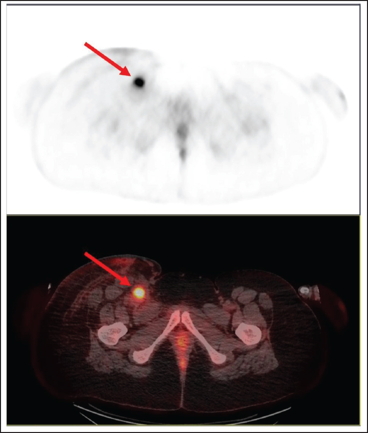
Figure 2. Axial T2W image (A) and axial post-contrast T1W image (B) of right upper thigh showing subcutaneous and intramuscular plane enhancing lesions (yellow arrows) encasing vessels (red arrow).
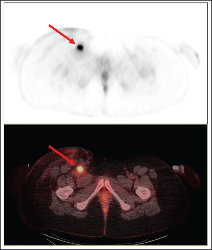
Figure 4. Multiple mildly metabolically active soft tissue density nodular lesions (red arrow) seen in right inguinofemoral region muscle and subcutaneous plane and associated with subcutaneous fat strandings/edema in right limb (recurrence) with multiple lymph nodes.
Timeline
| Time Point (Day) | Clinical Course and Interventions |
|---|---|
| Day 0 (Presentation) | The patient presented with swelling and pain in the right inguinal region |
| Day 2-7 | Diagnostic evaluation performed(laboratory investigations and radiological imaging.) |
| Day 15 | Underwent surgery |
| Day 70 | Post-operative assessment and investigations completed. |
| Day 82 | Full clinical recovery documented. |
| Day 85 | Adjuvant chemotherapy initiated. |
| Day 175 | Completion of chemotherapy (final cycle administered). |
| Day 348 | recurrence of disease seen. |
Discussion and Conclusions
Synovial sarcoma (SS) is a rare and aggressive subtype of soft tissue sarcoma (STS), comprising only 5%-10% of all STSs [1,2]. It is a malignant tumour of mesenchymal origin exhibiting variable degrees of epithelial differentiation [2]. Despite its name, SS is in fact a misnomer as it does not arise from synovial tissue [3-5] and is driven by a specific SS18-SSX fusion oncogene resulting from the t(X;18)(p11.2;q11.2) translocation [1-16].
The intravascular presentation of SS (IVSS) is an exceptionally rare entity. To the best of our knowledge, only 14 cases of IVSS [3-6,8-15] have been reported in the global literature, making this the 15th report worldwide.
IVSS is highly aggressive and characterised by tumour growth within vascular structures. Its prognosis is influenced by several factors, including tumour size, depth, anatomical location, invasion into surrounding tissues, and most importantly, achieving a complete surgical resection [1,5,10,13]. The intravascular location of the tumour contributes to a significantly higher risk of distant metastasis [15]. Our patient’s clinical course underscores this aggressive nature, where local recurrence was detected only 5 months after completion of CT.
In our case, the clinical presentation and initial imaging were suggestive of a DVT. However, it was actually the mass exerting significant extrinsic compressive effects and luminal obstruction of the vein. This resulted in an absence of definite colour flow or a venous spectral waveform, which was initially misinterpreted as a DVT. It can also present as a pulmonary embolism (PE) [4,13], superior vena cava (SVC) syndrome/obstruction (SVCO) [3], pleural effusion [14], and other thromboembolic events depending upon the location of the mass.
This is well illustrated in the case reported by Tong et al. [3], where the IVSS involved the SVC, presenting clinically as a cervical mass with features of SVC syndrome. Similar obstacles have been documented by Schreiner et al. [4] and Schoneveld et al. [13], who reported cases of IVSS initially mimicking common thromboembolic events. This creates a critical diagnostic pitfall where we can mistake a malignant intravascular process for a primary DVT.
To navigate this, a multimodal approach including clinical assessment, imaging, histopathology, IHC, and molecular studies [3-5,10,14,15] is required. Differentiating tumour thrombus from bland thrombus is key, and while Doppler USG is first-line for DVT, advanced imaging is crucial when atypical or malignant features such as internal vascularity and calcific foci are present, as seen in our case [12]. CT venography identified the enhancing mass, and FDG PET CT (Figure 3) confirmed the strong suspicion for a malignant intravascular process. The above-mentioned radiological red flags, along with atypical thrombotic presentations, particularly in young patients with a failure to respond to anticoagulation, should prompt thorough assessment and consideration of intravascular neoplasms to help prevent delay in diagnosis [3,4,10,12].
In our case, histopathological examination and IHC played a crucial role in the diagnostic workup, where the strong expression of SS18 helped in making the diagnosis of SS [17]. SS18 expression is a positive immunoexpression of the SS18::SSX fusion-specific antibody, which is highly specific for SS [18]. IHC also helped rule out neural origin tumours [7], mesothelioma, sex cord-stromal tumours, and carcinomas of the female genital tract [17]. Our case demonstrated significant downregulation of INI1 (SMARCB1) expression. In SS, several studies have reported reduced or mosaic loss of SMARCB1/INI1 protein expression in a large number of cases. This reduction is not due to gene deletion but is attributed to post-transcriptional regulatory mechanisms affecting protein expression within tumour cells. Therefore, the observed downregulation in our sample supports our diagnosis [19]. This IHC profile is consistent with previously published reports. Given the significant morphologic heterogeneity exhibited by SS [2], the use of molecular techniques remains critical for confirming the diagnosis [1-5,10,13-15].
The diagnosis in this case was definitively confirmed by the detection of the SYT-SSX1 gene fusion by FISH, a characteristic molecular alteration unique to this tumour type [1,2,4,5,14]. It is seen approximately in 95% of all cases [4], making it an exceptionally reliable diagnostic marker and also believed to be the cause [1]. This chromosomal reciprocal translocation [5] forms from the fusion of the SS18 gene on chromosome 18 with an SSX gene (SSX1 or SSX2) on the X chromosome [1,2,10,14].
The management of IVSS remains undefined due to the lack of standardized treatment guidelines and presents a significant predicament. Surgical resection remains the cornerstone of treatment [5,13], RT playing a crucial adjunctive role in improving prognosis [13] and reducing the risk of local recurrence. However, the role of CT remains controversial [5,13].
Several reports highlight the need for adapted strategies in IVSS due to its intravascular location and technical surgical challenges. Schreiner et al. [4] utilized a combination of neoadjuvant CT (doxorubicin and ifosfamide) and RT before surgical resection [4]. Maekura et al. [14] reported a case of unresectable pulmonary artery IVSS, in which combined CTRT was utilized as a disease-controlling strategy. Jonathan et al. [10] also reported a case wherein neoadjuvant RT was employed before radical en bloc excision of the tumour, followed by combined arterial and venous reconstruction, resulting in 2 years of disease-free survival. These approaches suggest that in cases where the tumour is large or located in anatomically critical regions, upfront surgery may not be feasible. Hence, preoperative RT or combined chemoradiotherapy (CTRT) may be employed in such cases.
In our case, the patient underwent surgery and was pathologically staged as pT2N0Mx with clear margins. Margin status is recognized as one of the most significant prognostic indicators for local recurrence in STS [16]. Achieving an R0 resection is therefore of paramount importance [5,16], as conservative surgical approaches have been associated with recurrence rates as high as 70% [5]. Additionally, tumour size has also been shown to correlate strongly with both local recurrence and systemic outcomes [10]. Lesions measuring greater than 5 cm in diameter are associated with a significantly increased risk of recurrence [13].
Postoperatively, our patient received five cycles of adjuvant CT with ifosfamide and doxorubicin. Although the role of adjuvant CT in synovial sarcoma remains debated [1,5,13], it was considered appropriate in this case due to the tumour’s size (>5 cm), high-grade histology, localized disease, negative lymph nodes, and the patient’s young age. RT was not administered given the anatomical location and the elevated risk of chronic lymphedema, particularly following extensive lymph node dissection and vascular involvement.
Management strategies are typically extrapolated from conventional synovial sarcoma, and reported cases show variability in therapeutic approaches [4-6,8,10,13-15], reflecting the need for an individualized nature of decision making based on tumour location, size, and extent of vascular involvement. A notable clinical feature is its tendency for late recurrence, frequently occurring beyond 5 years after initial diagnosis and treatment [2,6,16], underscoring the necessity for prolonged follow-up.
What sets our case apart is the unusually early locoregional recurrence within 5 months of receiving adjuvant CT (Figures 1–3) and within a year of surgical resection. This suggests that IVSS may have a more aggressive biology, possibly influenced by factors associated with the tumour microenvironment and mechanisms of vascular spread, and this is a major learning point showing the necessity for stringent monitoring.
A notable limitation of this case is the relatively short surveillance period, as the patient was lost to follow-up shortly after recurrence was documented. Consequently, any further interventions could not be carried out, and the ultimate clinical outcome could not be further evaluated.
Conclusion
In conclusion, this case represents a rare intravascular presentation of synovial sarcoma in a young woman, masquerading as DVT, a critical diagnostic pitfall. It highlights the critical importance of maintaining a high index of suspicion in atypical presentations with unprovoked thrombotic events and employing advanced imaging modalities for differentiation. Furthermore, the early locoregional recurrence despite optimal management emphasizes the aggressive nature of this disease. Given the limited literature, there is an unmet need for enhanced research into tumour biology, recurrence mechanisms, advanced molecular testing, and larger cohort-based studies to better define disease behaviour, refine prognostic markers, and guide tailored management approaches in the future.
What is new?
Intravascular synovial sarcoma (14 cases documented worldwide) is a rare condition whose management remains undefined, and surgical resection remains the standard treatment. Radiation has an adjunctive role in improving prognosis, and the role of chemotherapy remains controversial. Management strategies are typically extrapolated from conventional synovial sarcoma. It has a tendency for late recurrence and is aggressive in nature.
This case (15th case) demonstrates the early locoregional recurrence after an R0 surgical resection, despite negative lymph nodes, clear margins, and receiving adjuvant CT. This early relapse suggests that IVSS may have a more aggressive biology, possibly influenced by factors associated with the tumour microenvironment and mechanisms of vascular spread.
List of Abbreviations
| CT | Chemotherapy |
| DVT | Deep vein thrombosis |
| IHC | Immunohistochemistry |
| IVSS | Intravascular synovial sarcoma |
| MRI | Magnetic resonance imaging |
| PET-CT | Positron Emission Tomography Computed Tomography |
| PE | Pulmonary embolism |
| RT | Radiotherapy |
| STS | Soft tissue sarcoma |
| SVC | Superior vena cava |
| SVCO | Superior vena cava obstruction |
| SS | Synovial sarcoma |
| USG | Ultrasonography |
Conflict of interest
The authors declare that they have no conflict of interest regarding the publication of this case report.
Funding
No funding was received for the preparation of this case report.
Consent for publication
Consent obtained directly from patient.
Ethics approval and consent to participate
Not applicable
Availability of data and materials
All data used in this case report are derived from the sources cited in the reference section below. No additional datasets were generated or analysed during the preparation of this manuscript.
Author details
Ritwika Choudhury1, Bhumi Agarwal2, Ambarish Satwik3, Mansi Sharma2, Abhinav Dewan2, Sabeena K. Choudhary2, Sunil Pasricha4, Himanshu Rohela5, Ullas Batra2
- Department of Research (Medical Oncology Unit 5), Rajiv Gandhi Cancer Institute & Research Centre, New Delhi, India
- Department of Medical Oncology, Rajiv Gandhi Cancer Institute & Research Centre, New Delhi, India
- Department of Vascular and Endovascular Surgery, Sir Ganga Ram Hospital, Delhi, India
- Department of Histopathology, Rajiv Gandhi Cancer Institute and Research Centre, New Delhi, India
- Department of Orthopaedic Oncology, Rajiv Gandhi Cancer Institute and Research Centre, New Delhi, India
References
- Aytekin MN, Öztürk R, Amer K, Yapar A. Epidemiology, incidence, and survival of synovial sarcoma subtypes: sEER database analysis. J Orthop Surg (Hong Kong). 2020;28(2): 2309499020936009. https://doi.org/10.1177/2309499020936009
- Thway K, Fisher C. Synovial sarcoma: defining features and diagnostic evolution. Ann Diagnostic Pathol. 2014;18(6):369–80. https://doi.org/10.1016/j.anndiagpath.2014.09.002
- Tong GX, Goldblum JR, Qiu W, Skacel M, Downs-Kelly E, Contractor S, et al. Primary intravascular synovial sarcoma: a disease of young adult women? Report of a case diagnosed by aspiration biopsy and review of the literature. Diagn Cytopathol. 2006;34(12):834–8. https://doi.org/10.1002/dc.20574
- Schreiner M, Sanad W, Pfitzner BM, Baumann G, Knebel F. A primary intravascular synovial sarcoma causing deep-vein thrombosis and pulmonary embolism in a 20-year-old woman. Curr Oncol. 2015;22(5):e387–90. https://doi.org/10.3747/co.22.2315
- Wise KB, Said SM, Clark CJ, Okuno SH, Shah SS, Park SJ, et al. Resection of a giant primary synovial sarcoma of the inferior vena cava extending into the right atrium with caval reconstruction under cardiopulmonary bypass and circulatory arrest. Perspect Vasc Surg. 2012;24(2):95–101. https://doi.org/10.1177/1531003512468035
- Miettinen M, Santavirta S, Slätis P. Intravascular synovial sarcoma. Hum Pathol. 1987;18(10):1075–7. https://doi.org/10.1016/S0046-8177(87)80227-7
- Golouh R, Vuzevski V, Bracko M, Van Der Heul RO, Cervek J. Synovial sarcoma: a clinicopathological study of 36 cases. J Surg Oncol. 1990;45(1):20–8. https://doi.org/10.1002/jso.2930450106
- Robertson NJ, Halawa MH, Smith ME. Intravascular synovial sarcoma. J Clin Pathol. 1998;51(2):172–3. https://doi.org/10.1136/jcp.51.2.172
- Shaw GR, Lais CJ. Fatal intravascular synovial sarcoma in a 31-year-old woman. Hum Pathol. 1993;24(7):809–10. https://doi.org/10.1016/0046-8177(93)90021-8
- White JS, Medlicott SAC, Brown H, Moore R, Temple W. Intravascular synovial sarcoma of the external iliac vein and reconstruction with the superficial femoral vein. J Vascular Surg. 2005;42(2):365–7. https://doi.org/10.1016/j.jvs.2005.03.059
- Tuncer ON, Erbasan O, Golbasi I. Primary intravascular synovial sarcoma: case report. Heart Surg Forum. 2012;15(5):E297–299. https://doi.org/10.1532/HSF98.20111159
- Yan H, Zhou C, Yan F, Wen X, Luo Y. Case 290: intravascular Cystic Synovial Sarcoma. Radiology. 2021;290(3):730–5. https://doi.org/10.1148/radiol.2021192863
- Schoneveld JM, Debing E, Verfaillie G, Geers C, Van Den Brande P. Deep venous thrombosis and pulmonary embolism caused by an intravascular synovial sarcoma of the common femoral vein. Vasc Endovascular Surg. 2012;46(8):693–5. https://doi.org/10.1177/1538574412460770
- Maekura T, Shimizu S, Kawaguchi T, Morimoto M, Okuma T, Matsui H, et al. Intravascular synovial sarcoma of the pulmonary artery with massive pleural effusion: report of a case with a favorable response to Ifosfamide chemotherapy and palliative radiation therapy. Intern Med. 2015;54(9):1095–8. https://doi.org/10.2169/internalmedicine.54.3624
- Chicas-Sett R, Farga-Albiol D, Collado E, Pacheco A, Zac C, Diaz R, et al. Intravascular Biphasic Synovial Sarcoma: the Beneficial Role of Adjuvant Treatment Approach in the Pre-metastatic Stage. Cureus. 2016;8(4):e572. https://doi.org/10.7759/cureus.572
- Italiano A, Penel N, Robin YM, Bui B, Le Cesne A, Piperno-Neumann S, et al. Neo/adjuvant chemotherapy does not improve outcome in resected primary synovial sarcoma: a study of the French Sarcoma Group. Ann Oncol. 2009;20(3):425–30. https://doi.org/10.1093/annonc/mdn678
- Lasota J, Chłopek M, Kaczorowski M, Natálie K, Ryś J, Kopczyński J, et al. Utility of Immunohistochemistry With Antibodies to SS18-SSX Chimeric Proteins and C-Terminus of SSX Protein for Synovial Sarcoma Differential Diagnosis. Am J Surg Pathol. 2024;48(1):97–105. https://doi.org/10.1097/PAS.0000000000002144
- Zaborowski M, Vargas AC, Pulvers J, Clarkson A, De Guzman D, Sioson L, et al. When used together SS18-SSX fusion-specific and SSX C-terminus immunohistochemistry are highly specific and sensitive for the diagnosis of synovial sarcoma and can replace FISH or molecular testing in most cases. Histopathology. 2020;77(4):588–600. https://doi.org/10.1111/his.14190
- Kohashi K, Oda Y, Yamamoto H, Tamiya S, Matono H, Iwamoto Y, et al. Reduced expression of SMARCB1/INI1 protein in synovial sarcoma. Mod Pathol. 2010;23(7):981–90. https://doi.org/10.1038/modpathol.2010.71
Summary of the case
| 1 | Patient (gender, age) | 22 years, Female |
| 2 | Final diagnosis | Intravascular synovial sarcoma arising from the right external iliac vein and common femoral vein. |
| 3 | Symptoms | Swelling and pain in the right inguinal region |
| 4 | Medications | Ifosfamide and adriamycin regimen |
| 5 | Clinical procedure | Surgery followed by chemotherapy |
| 6 | Specialty | Oncology |
Keywords: 15th case, intravascular synovial sarcoma, deep vein thrombosis, thromboembolic events, diagnostic challenge.


Publication History
Received: March 09, 2026
Revised: April 25, 2026
Accepted: April 30, 2026
Published: July 25, 2026
Authors
Ritwika Choudhury
Department of Research (Medical Oncology Unit 5), Rajiv Gandhi Cancer Institute & Research Centre, New Delhi, India.
 ORCID
Bhumi Agarwal
Department of Medical Oncology, Rajiv Gandhi Cancer Institute & Research Centre, New Delhi, India.
Ambarish Satwik
Department of Vascular and Endovascular Surgery, Sir Ganga Ram Hospital, Delhi, India.
Mansi Sharma
Department of Medical Oncology, Rajiv Gandhi Cancer Institute & Research Centre, New Delhi, India.
Abhinav Dewan
Department of Medical Oncology, Rajiv Gandhi Cancer Institute & Research Centre, New Delhi, India.
Sabeena K Choudhary
Department of Medical Oncology, Rajiv Gandhi Cancer Institute & Research Centre, New Delhi, India.
Sunil Pasricha
Department of Histopathology, Rajiv Gandhi Cancer Institute and Research Centre, New Delhi, India.
Himanshu Rohela
Department of Orthopaedic Oncology, Rajiv Gandhi Cancer Institute and Research Centre, New Delhi, India.
Ullas Batra
Department of Medical Oncology, Rajiv Gandhi Cancer Institute & Research Centre, New Delhi, India.